
Pelvic Organs Female Diagram Abdominal Anatomy Chart Female Anatomy
The diaphragm marks the top of the abdomen and the horizontal line at the level of the top of the pelvis marks the bottom. Connective tissue called the mesentery holds the abdominal organs together. Several large blood vessels travel through the abdomen.
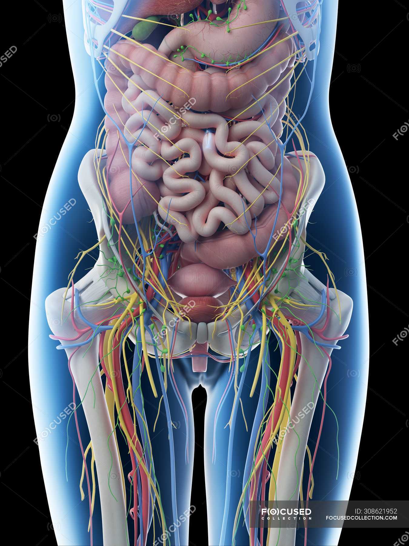
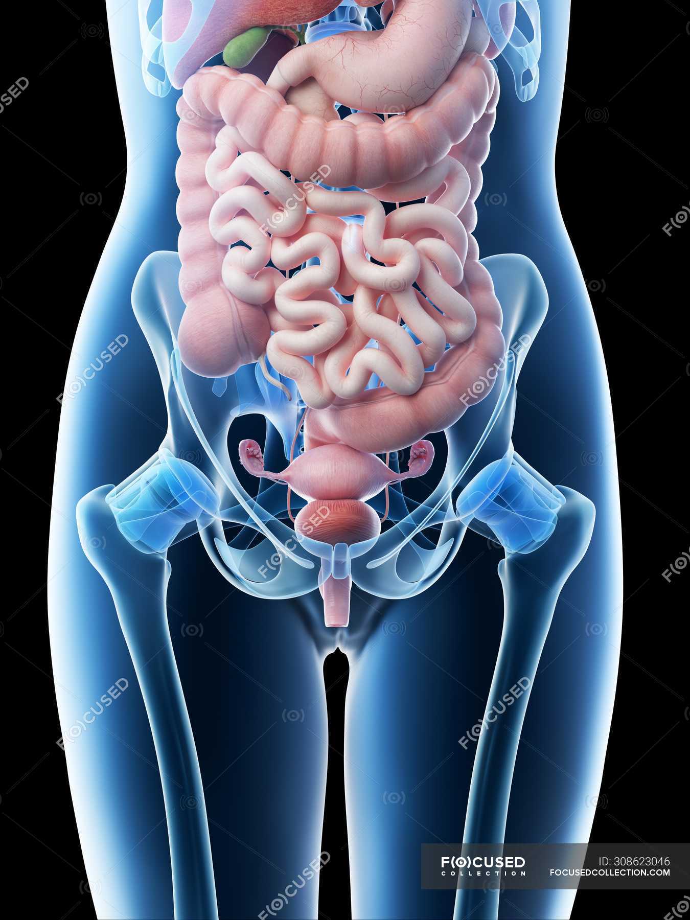
Female abdominal anatomy and internal organs, computer illustration
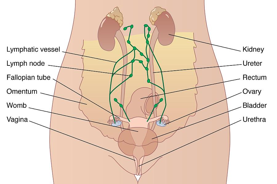
The bladder, also known as the urinary bladder, is an expandable, muscular sac that stores urine. When signaled, the bladder releases urine into the urethra, a tube that carries it out of the body..

Female Abdominal Anatomy TrialExhibits Inc.
The abdomen is the part of the body that contains all of the structures between the thorax (chest) and the pelvis, and is separated from the thorax via the diaphragm. The region occupied by the abdomen is called the abdominal cavity, and is enclosed by the abdominal muscles at front and to the sides, and by part of the vertebral column at the back.

Female Anatomy Upper Body Stock Photo Download Image Now iStock
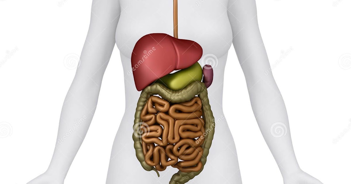
ISSN 2534-5079. This e-Anatomy illustrates the gross anatomy of the digestive system. We focused especially on the diagrams of the abdominal digestive system (oesophagus is described on the modules about the thorax and oral cavity/pharynx on the ENT modules). 84 anatomical diagrams and histological images with over 300 labeled anatomical parts.
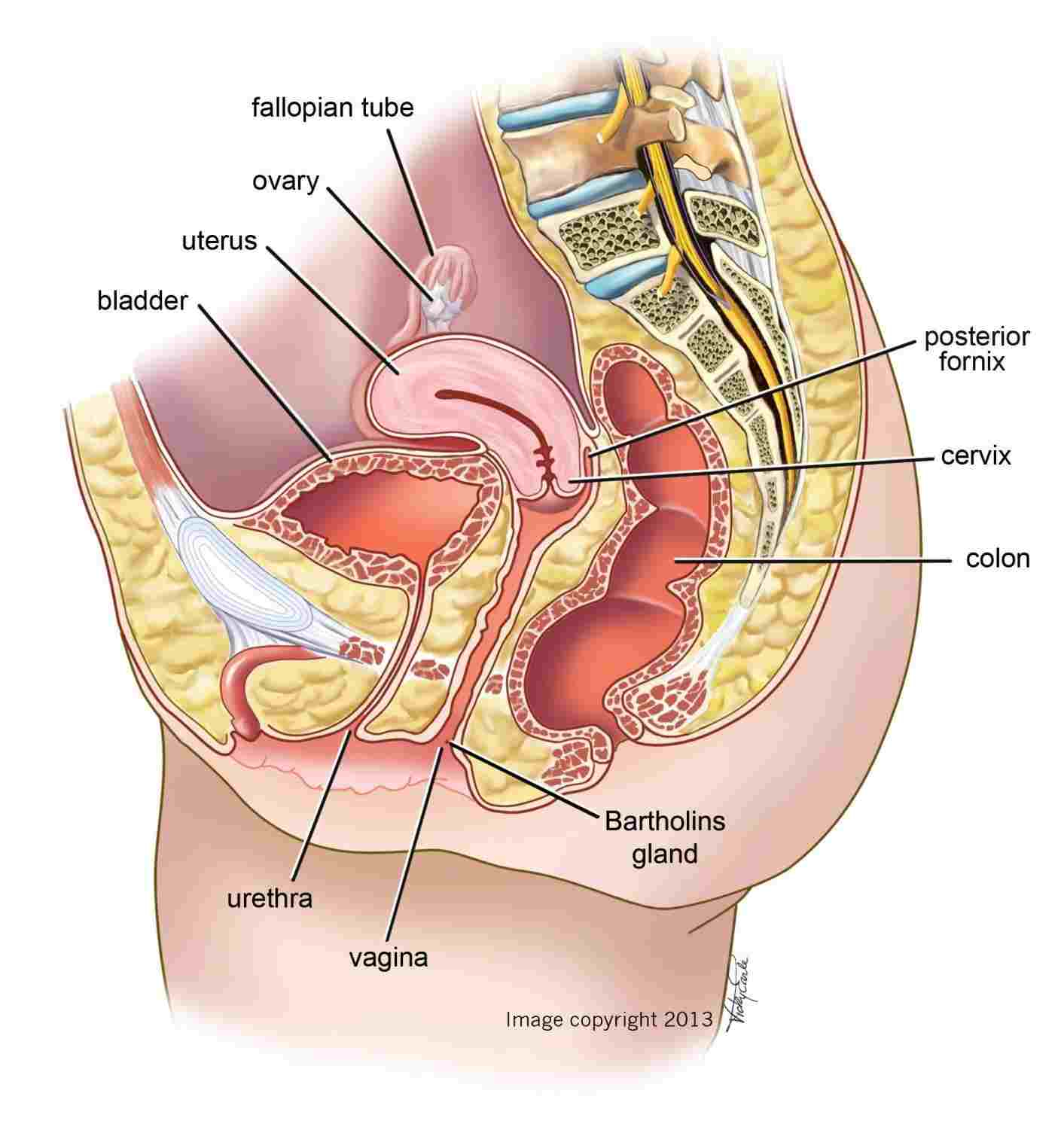
Diagram Of Abdominal Organs exatin.info
Vulva Female reproductive organs are very different to those of males. The vulva refers to the external parts of a female's genitals. It consists of several parts, including the labia majora,.

Abdominal Anatomy Pictures Female Female Human Body Organs Diagram
Uterus. Also called the womb, the uterus is a hollow, pear-shaped organ located in a woman's lower abdomen, between the bladder and the rectum. Ovaries. Two female reproductive organs located in the pelvis. Fallopian tubes. Carry eggs from the ovaries to the uterus. Cervix.

Human Anatomy Abdomen Anatomy Pinterest Human anatomy
Cite this Item Add to Collection This medical illustration depicts a mid-sagittal view of the normal anatomy of the female abdomen and pelvis. Labeled structures include the large bowel (colon or large intestine), umbilicus, small intestine, ovary, fallopian tube, uterus and bladder. Variations
Abdomen AnatomyFemale Female Abdominal Anatomy Illustration Stock
Abdominal diagram 1013 Abdominal diagram 1014 Abdominal diagram 1018 Abdominal diagram 1022 Abdominal diagram 1030 Abdominal diagram 1069 Abdominal diagram 1153 Abdominal diagram 1375 Abdominal diagram 1424 Abdominal diagram 1564 This article is about Anatomy Of The Female Abdomen And Pelvis, Cut away View.
Abdominal Anatomy Pictures Female Female abdominal anatomy, computer
Quizzes Abdomen Peritoneum and peritoneal cavity Stomach Spleen Pancreas Liver and gallbladder Small intestine Large intestine Kidneys, ureters and adrenal glands Pelvis Perineum Urinary bladder and urethra Female reproductive organs Male reproductive organs Blood vessels Innervation Lymphatics Sources Related articles Abdomen and pelvis
Anatomy Of The Female Abdomen And Pelvis, Cut away View Healthiack
The main bones in the abdominal region are the ribs. The rib cage protects vital internal organs. There are 12 pairs of ribs and they attach to the spine. There are seven upper ribs, known as.

de Female Human Anatomy Organs Diagram mar webmds abdomen anatomy page
Diagram External Internal Breast Anatomy Functions Female anatomy includes the internal and external structures of the reproductive and urinary systems. Reproductive anatomy plays a role in sexual pleasure, getting pregnant, and breastfeeding. The urinary system helps rid the body of toxins through urination (peeing).

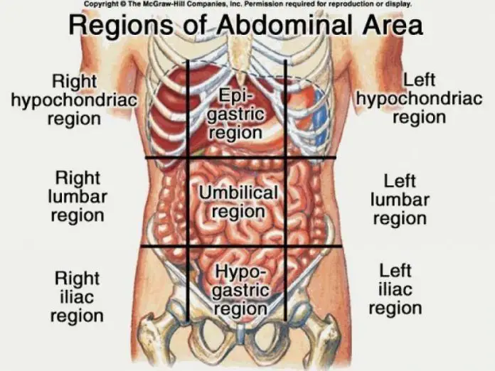
Abdominal Regions and Associated Pain
Anatomy atlas of the female pelvis: 101 labeled illustrations of the female genital system (ovaries, uterine tubes, uterus, vagina, vulva, clitoris) and pelvic cavity (bladder, rectum, pelvic diaphragm, perineum with innervation and blood supply). Tome 2 : Thorax, coeur, abdomen et pelvis. Torsten B. Möller - Emil Reif. Paru le : 06/2014.
Anatomy Of The Female Abdomen And Pelvis, Cut away View Healthiack
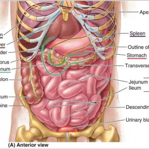
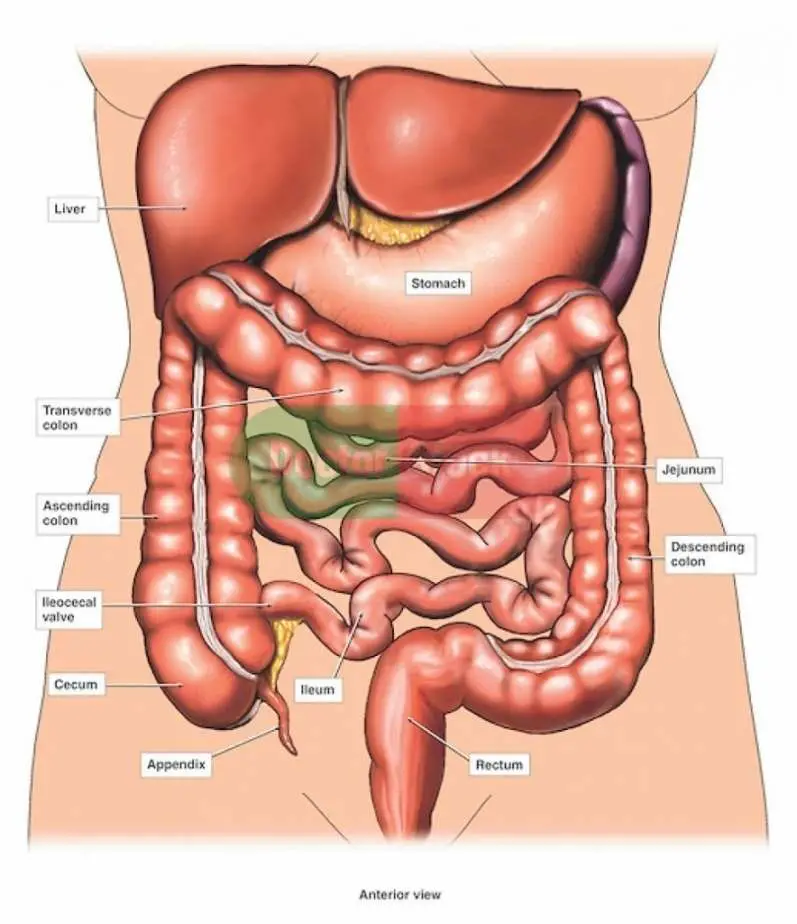
1. Anterior view: anatomy of female abdomen and pelvis: skin 2. Anterior view: anatomy of female abdomen and pelvis: muscles of anterior abdomen wall 3. Anterior view: anatomy of female abdomen and pelvis: stomach and omentum 4. Anterior view: anatomy of female abdomen and pelvis: small bowel and colon 5.
Anatomy Of The Female Abdomen And Pelvis, Cut away View
The abdomen (colloquially called the belly, tummy, midriff, tucky or stomach) is the part of the body between the thorax (chest) and pelvis, in humans and in other vertebrates. The abdomen is the front part of the abdominal segment of the torso. The area occupied by the abdomen is called the abdominal cavity.

Female Abdominal Organs Diagram / Instant Anatomy Abdomen Areas
Pulled or strained abdominal muscles Cirrhosis of the liver Colon cancer Last medically reviewed on October 23, 2014 The muscles of the abdomen protect vital organs underneath and provide.
Female Abdominal Anatomy Pictures / Stock Images Female Abdominal
Browse 617 female anatomy diagram photos and images available, or start a new search to explore more photos and images. of 11 NEXT Browse Getty Images' premium collection of high-quality, authentic Female Anatomy Diagram stock photos, royalty-free images, and pictures.